My camera has turned into a microscope
Warning: highly detailed images, please exercise patience.
Notice: there are several typographical errors on the images. It is best to ignore all 0.20 markings.
After some optical black magic with the aid of Adrian and Diana's Canon 28-80mm EF lenses, I have been able to turn my Panasonic FZ30 into a microscope capable of resolving details about 760 nm per pixel.
Interestingly, the wavelength of red light is 700 nm, and I can see minor diffraction effects due to the wave nature of propagating light.
Right, let's get to the photographs...
Click here for large size image
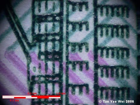
The scale is corresponds to a length of 1.0 mm, subdivided into 0.5 mm and 0.2 mm sections.
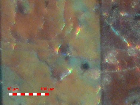
This is an image of a polymer RM 5 banknote. It is part of the Petronas Twin Towers, where the diagonal line on the left is the bottom of the right supporting-strut of the sky bridge connecting the towers.
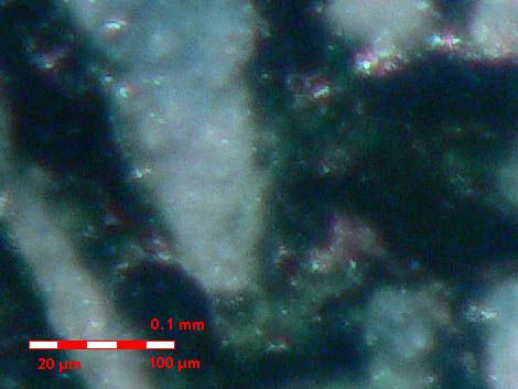
The scale is corresponds to a length of 0.1 mm, subdivided into 20 μm (micrometres, microns) sections.
Zooming in to the full sized photo (100% crop), one can actually see the thickness variations on the black-ink film.
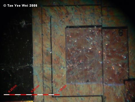
The next image is of the holographic strip on a paper RM 50 banknote. The holographic strip includes patterns that look like 4 little squares arranged around the corners of a slightly larger square.
Click here for large size image
The scale is corresponds to a length of 1.0 mm, subdivided into 0.1 mm sections.
The subsequent image is a slight close-up of a corner of those little squares. One can see that the zigzagging band of light around the square array is caused by what appears to be a groove carved into the metallic surface.
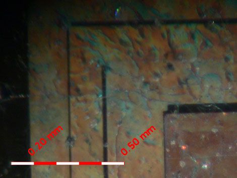
There is a typo on the image- the first number on the scale should read 0.1 mm, not 0.2 mm.
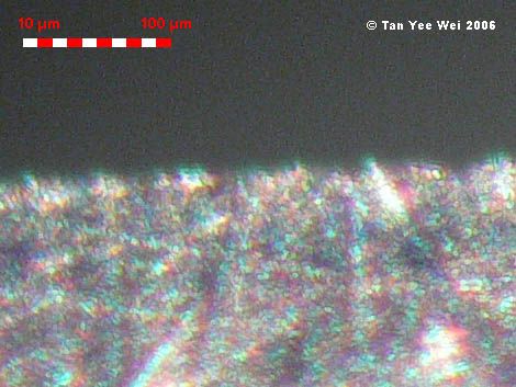
The following image is a 100% crop of the full-sized photo. Note the surface textures on the ‘flat’ regions of the holographic surface.
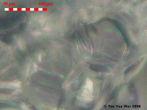
The scale is corresponds to a length of 0.1 mm, subdivided into 10 μm (micrometres, microns) sections.
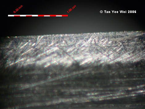
This is a photograph of the freshly sharpened cutting edge of a kitchen knife.
Click here for large size image
At maximum magnification, the diffraction effects of light are visible, particularly at the knife’s edge. Faint diffraction patterns are visible against the dark background.
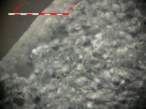
A thinly sliced cross section of bak choy (白菜,a vegetable)stem. The visible granular structure appears to be composed of individual cells, although that suspicion cannot be confirmed.
Click here for large size image
This is a 100% crop of the previous image. If the individual particles are indeed cells, then I would suggest that the voids within each cell are the vacuoles (air pocket present in certain plant cells).
Click here for large size image
Put a ruler up to your monitor to measure how long the 0.1 mm sections appears on your monitor. On mine, it is a surprisingly convenient 40 mm, yielding a magnification of 400x. Better than most optical microscopes, I’ll say.
Photographs
Interesting reads
Mathematics & Applied Sciences
Notice: there are several typographical errors on the images. It is best to ignore all 0.20 markings.
After some optical black magic with the aid of Adrian and Diana's Canon 28-80mm EF lenses, I have been able to turn my Panasonic FZ30 into a microscope capable of resolving details about 760 nm per pixel.
Interestingly, the wavelength of red light is 700 nm, and I can see minor diffraction effects due to the wave nature of propagating light.
Right, let's get to the photographs...
Click here for large size image
The scale is corresponds to a length of 1.0 mm, subdivided into 0.5 mm and 0.2 mm sections.
This is an image of a polymer RM 5 banknote. It is part of the Petronas Twin Towers, where the diagonal line on the left is the bottom of the right supporting-strut of the sky bridge connecting the towers.
The scale is corresponds to a length of 0.1 mm, subdivided into 20 μm (micrometres, microns) sections.
Zooming in to the full sized photo (100% crop), one can actually see the thickness variations on the black-ink film.
The next image is of the holographic strip on a paper RM 50 banknote. The holographic strip includes patterns that look like 4 little squares arranged around the corners of a slightly larger square.
Click here for large size image
The scale is corresponds to a length of 1.0 mm, subdivided into 0.1 mm sections.
The subsequent image is a slight close-up of a corner of those little squares. One can see that the zigzagging band of light around the square array is caused by what appears to be a groove carved into the metallic surface.
There is a typo on the image- the first number on the scale should read 0.1 mm, not 0.2 mm.
The following image is a 100% crop of the full-sized photo. Note the surface textures on the ‘flat’ regions of the holographic surface.
The scale is corresponds to a length of 0.1 mm, subdivided into 10 μm (micrometres, microns) sections.
This is a photograph of the freshly sharpened cutting edge of a kitchen knife.
Click here for large size image
At maximum magnification, the diffraction effects of light are visible, particularly at the knife’s edge. Faint diffraction patterns are visible against the dark background.
A thinly sliced cross section of bak choy (白菜,a vegetable)stem. The visible granular structure appears to be composed of individual cells, although that suspicion cannot be confirmed.
Click here for large size image
This is a 100% crop of the previous image. If the individual particles are indeed cells, then I would suggest that the voids within each cell are the vacuoles (air pocket present in certain plant cells).
Click here for large size image
Put a ruler up to your monitor to measure how long the 0.1 mm sections appears on your monitor. On mine, it is a surprisingly convenient 40 mm, yielding a magnification of 400x. Better than most optical microscopes, I’ll say.
Photographs
Interesting reads
Mathematics & Applied Sciences
Labels: microscopy, photographic equipment, photography
posted by Tan Yee Wei at 7/28/2006 01:29:00 am
![]()
![]()

<< Home